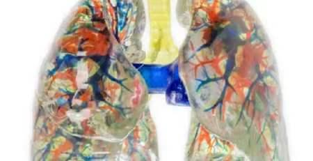
Medical academies face a persistent deficit in anatomical training resources. Traditional formalin-fixed cadavers remain scarce, and sourcing biological specimens entails complex logistics. Educational facilities require scalable alternatives that maintain rigorous clinical standards. Modern industrial processes now provide a robust solution. Engineers construct physical replicas using high-precision digital human datasets.
These 1:1 physical models deliver exact anatomical geometry. The process bridges the gap between a virtual dissection table and tactile surgical training. Students interact with physical specimens that perfectly mirror complex biological structures. This technological workflow guarantees standardized educational materials across medical institutions.
Integrating Data from the virtual dissection table
Translating biological structures into physical forms demands extreme data precision. The workflow starts within a digital environment. Medical students and instructors frequently utilize a virtual dissection table to isolate specific organ systems. This digital platform relies on a vast dataset. The dataset includes original sectional data, refined segmentation structures, and complete three-dimensional geometric models. To guarantee visual and structural fidelity, technicians extract volume data voxels directly from the surface of these digital structures.
The source data achieves a voxel size of 0.0384 mm × 0.0384 mm × 0.1 mm. This extreme density captures the minutiae of human biology. Engineers use these extracted voxels to generate a texture map. They apply this map directly to the corresponding geometric model. This precise digital mapping ensures the final output looks identical to a real anatomical specimen. The foundational dataset covers all vital structures. These include bone, muscle, blood vessels, nerves, and ligaments. Medical instructors isolate these structures digitally before sending the finalized files to the physical production queue.
Multi-Channel Hardware and Material Specifications
Standard commercial printers fail to replicate human tissue complexity. Medical-grade reproduction requires specialized industrial machinery. Facilities utilize full-color, multi-material 3D printers built specifically for medical simulation. These machines combine 3D inkjet printing technology with advanced light curing mechanisms. According to the 3D+Printing+Models+Brochure, the hardware features 12 distinct material channels, allowing the simultaneous deposition of diverse chemical compounds during a single print cycle.
Printing complex organs requires varied physical properties. The hardware supports multiple material configurations. Technicians can execute full-color hard printing or soft-hard composite printing. They can also perform full-color soft printing using environmentally friendly materials. Available materials range from rubber-like polymers to rigid plastics. The system also utilizes transparent packaging materials alongside opaque resins.
The print mechanism contains exactly 3840 piezoelectric spray holes. These nozzles operate via proprietary high-frequency spray curing algorithms. This highly integrated system achieves a rapid spray volume of 4 liters per hour. The manufacturing cycle utilizes efficient post-processing equipment. This auxiliary machinery eliminates the need for secondary curing. Academies receive highly accurate printed models on accelerated production timelines.
Validating Physical Fidelity and Tactile Resistance
Digital models provide excellent spatial orientation. However, surgeons require physical tactile feedback. Tactile resistance is critical for surgical planning and muscle memory. The resulting 3D printed models deliver a physical weight comparable to actual biological organs. They also mimic the soft and hard tactile properties of real anatomical specimens. Instructors use these physical models to demonstrate complex surgical pathways safely.
The printed specimens undergo rigorous validation against formalin-fixed cadavers. Medical professionals compare the digital models to the physical outputs. Minor variations in hue may exist between the screen and the physical print. However, the geometric morphology remains flawless. There is no significant difference in anatomical details, texture features, or overall physical scale.
Surgeons demand extreme accuracy when practicing intricate procedures. A skull model requires a rigid exterior, while internal cerebral arteries require flexible soft-tissue simulation. The multi-material printer constructs these delicate pathways inside a transparent supporting matrix. During the print cycle, the machine generates automatic support structures. These supports protect overhanging tissues and narrow internal cavities. Brain surgeons, hand surgeons, and clinical anatomists unanimously recognize the physical authenticity of these complex structural replicas.
Streamlining Institutional Procurement and Ecosystems
Upgrading an anatomy laboratory requires reliable technological ecosystems. Academic institutions must source both the digital datasets and the physical modeling hardware. Sourcing these components from isolated vendors often creates software integration issues. A unified digital-to-physical ecosystem eliminates these workflow bottlenecks.
Medical faculties require seamless transitions from digital exploration to physical interaction. A student might study cranial nerves digitally on a virtual dissection table. The next day, that same student holds the physical replica of that exact digital nerve pathway. This educational consistency reinforces spatial memory. It accelerates the overall learning curve for complex surgical disciplines.
Institutions deploying these systems report immediate improvements in training efficiency. Professors spend less time managing biological specimen preservation. They spend more time teaching applied surgical techniques. The integration of high-resolution digital data and rapid multi-material printing creates a highly scalable training environment. Medical directors looking to modernize their facilities can review the integrated digital anatomy solutions provided by DIGIHUMAN. Their comprehensive product lineup bridges the gap between software simulation and physical surgical practice.
Transforming Doctor-Patient Clinical Communication
These anatomical models also serve vital clinical communication purposes. Doctors frequently struggle to explain complex pathologies using flat diagnostic images. A two-dimensional scan lacks intuitive physical context. Patients often fail to grasp the severity or precise location of an internal issue.
Physical 3D printed models solve this communication barrier. A physician can hold a precise, 1:1 scale replica of a human heart. They can point to the exact location of a vascular blockage. Patients understand physical objects instantly. This tactile interaction improves patient consent rates. It builds immediate trust between the surgical team and the patient. Medical centers utilize these models to demystify complex preoperative consultations. The transition from abstract digital data to a physical educational tool benefits medical students and clinical patients equally.
